
What do cells in the nose, primary ciliary dyskinesia (PCD) and arrhythmia have in common? They were the topics of this year's winners from the annual research photo competition at Royal Brompton & Harefield NHS Foundation Trust.
With submissions from across both hospitals, this year’s crop of photos covered a wide ranging array of interpretations on the theme – what does research mean to you?
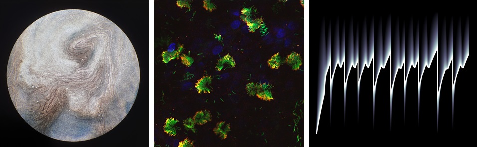
From left to right: First place ‘S’not what you think!’ by Ranjit Rai, second place ‘Fiery Bloom’ by Farheen Daudvohra and third place ‘Arrhythmia Peaks’ by Jack Allen.
On first look, the winning photo, titled “S’not what you think!” resembles the moon, or perhaps a planet, but according to the photographer it’s a microscopic close up of the cells that line the respiratory tract.
Ranjit Rai, a scientist working with the PCD team, explained the science behind her winning image:
“Goblet cells produce mucus in the airways and cilia (hair like projections on cells) are needed to beat back and forth, pushing this mucus along to clear the airways. In PCD the cilia beat in an abnormal way or don’t beat at all, leading to a build-up of mucus and increased risk of respiratory infections. The photo shows some of the beautiful streaks of mucus created by the cilia beating”.
And how did she manage to capture such a photo?
“I took the photo by placing the camera lens over the eyepiece of the microscope which shows you exactly what we see when we look for mucus and cilia. Growing these kind of cells in the lab is often a challenge, so it’s a lovely feeling to be able to share some of the beautiful patterns we see under the microscope with everyone.”
The judging panel, which included staff members from the Trust and a patient, scored the submissions based on a selection of criteria, including theme, composition and originality.
In second place we have “Fiery Bloom” by Farheen Daudvohra, another scientist from the paediatric PCD team. Her photo shows an immunofluorescent image of ciliated cells from a patient with PCD that has been dyed with different coloured stains. The blue stain highlights the cell nuclei, while the green structures depict the cilia.
And finally, in third place we have “Arrhythmia Peaks” from Jack Allen, a cardiovascular research associate working on MRI scans. Jack’s image shows a computer simulation of an MRI heart scan for a patient with an abnormal heart rhythm. Each shade of grey represents a particular tissue type, demonstrating how it changes during the scan (from left to right).
The winning photos will be used as part of the Trust's communication materials for research over the next year, so keep an eye out for them.
Take a look at last year's winners.
To find out more about our research please contact us.