
Specialists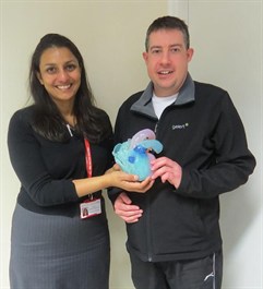 at Royal Brompton & Harefield NHS Foundation Trust are using 3D imaging and printing to create exact replicas of patients’ hearts to help them plan and carry out surgery.
The technique requires specialist cardiovascular magnetic resonance (CMR) imaging, which uses powerful magnets and radio waves to take detailed pictures of an individual’s heart.
Every CMR scan produces hundreds of images that are stored and displayed using advanced computer software, to create an exact three-dimensional digital replica of the human heart. This means experts can view the patient’s heart structure, including the muscles, chambers and valves, in a particularly lifelike way, to assess how well the heart is pumping and quickly diagnose a variety of problems, including:
- congenital heart disease
- wear and tear of heart valves
- any other damage to the heart.
New experts at the Trust are going one step further and using 3D printing to turn the digital replica into an actual exact model of a patient’s heart.
How is 3D imaging and printing used?
Dr Sonya Babu-Narayan, honorary consultant cardiologist and British Heart Foundation Imperial College clinical senior lecturer, is interested in the role of CMR in preventing and treating heart rhythm disturbances in patients born with congenital heart disease, as well as using 3D imaging during invasive heart procedures.
She says: “The availability of 3D modelling when preparing for and performing heart surgery or other cardiac procedures allows two really important things to happen.
“Firstly, surgeons and other clinical team members are able to better grasp how a patient’s heart is affected by their condition. This leads to better care and allows us to diagnose and repair conditions with less need for invasive diagnostic procedures.
“When we have an electronic 3D model of a patient’s heart we can store this on their record, an invaluable tool for follow up appointments.
“Secondly, a 3D model can be a huge help with the communication between the clinical team and the patient. A 3D visual representation of the heart is so much clearer than anything we could put in to words.
“We can even go one step further and get the model 3D printed. Coming in for surgery can be a worrying time for a patient, so we always like to fully explain what will happen to help put their mind at rest as much as possible.
“Seeing and handling an actual model of their heart can be so much more informative than two dimensional drawings or other images, especially for patients with complex congenital heart disease, both before and after interventions in the catheter laboratory or surgery.”
One of the other advantages of printing a 3D model of the human heart is the opportunity to improve training. Heart disease comes in many different forms and the more cardiologists can learn about the structure of hearts with these problems, the better they will be able to treat them.
Trainees can also practice on exact replicas of hearts, including ones with congenital heart diseases. The models used for this training are made of material that allows doctors to simulate a real-life situation. Some models even look like a real human heart under x-ray.
Groundbreaking innovation: scarring of the heart
The 3D programme at Royal Brompton, supported by biomedical engineers Materialise, recently did something that no other hospital in the world has done, which was to print hearts that also show regions of scarring.
Scarring, often caused by heart disease or surgery, is one of the reasons that patients may have life -threatening arrhythmia, a condition where the heart is beating too fast, slow or irregularly.
At Royal Brompton Hospital, using 3D modelling, consultants and surgeons can gain a better understanding of what kind of scarring causes different types of arrhythmia.
This research is invaluable for improving treatments for patients with arrhythmias in future. The aim is to prevent patients from requiring a defibrillator as a precaution and to only implant the lifesaving device when it is certain they need one.
The ability to view a 3D image of every unique heart in so much detail can also reduce the need for as many cardiac catheterisations and open heart surgery.
The Trust is continually looking to develop its 3D programme and the Royal Brompton & Harefield Hospital Charity is running a campaign to raise money towards a new 3D printer, advanced software and other vital resources.
A patient’s view
Jonathan Havre is a patient at Royal Brompton Hospital with repaired tetralogy of Fallot, one of the most common congenital heart conditions, which is characterised by four structural defects in the heart. As an adult, Jonathan’s CMR images were used for his electrophysiology procedure, to see how ‘at risk’ he was of rhythm disturbance related to postoperative scars in his heart.
The team used the CMR images to produce detailed prints of his heart muscle, including scarred areas, and were able to show him a 3D printed life size model of his own heart.
He said: “I found the model of the heart gave me a real understanding of what was happening. It really put me at ease knowing exactly what my heart looks like.
“It is sometimes hard to picture what I’m being told. With the 3D model I could hold my own heart, an experience I never thought I’d have.”
The future
3D techniques have been used in industry and in healthcare in many settings, but it is only more recently that the technology has been applied to the heart and vascular systems.
As 3D modelling develops, hospitals across the world will be able to use it to help with a variety of procedures and deliver improved patient care.
Application of these techniques should lead to reduced invasive procedures, improved medical learning and clearer communication between clinical staff and patients.
Dr Babu-Narayan is certain 3D technology will feature more prominently in the hospitals of the future.
“Across the world techniques are continually evolving, we are barely scratching the surface of the potential clinical uses for 3D imaging and printing. For example, there are now 3D bioprinters which use living cells as ink, giving an unrivalled representation of a select body part,” she said.
"Some healthcare settings are even testing virtual reality headsets. Holographic technology lets clinicians become fully immersed in the digital image of the scan away from a computer screen, possibly even in the operating theatre.”
"Finally, there is also interactive 3D technology that combines the tactile advantages of a printed model with the immense detail offered by the digital image. The future really is bright in this revolutionary field."
Find out more about Royal Brompton Hospital CMR unit.